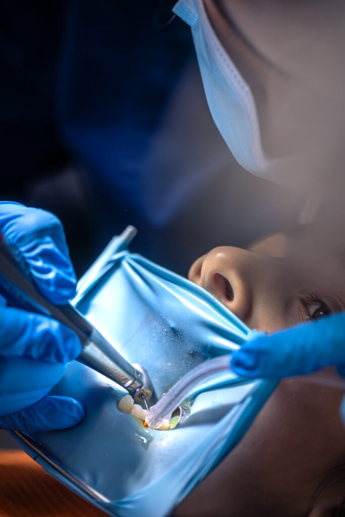
Microscopic Root Canal Treatment, also known as Micro-Endodontics, utilizes high-powered dental operating microscopes that provide up to 25x magnification and enhanced illumination. This advanced technology allows Dr. Siddartha to see the intricate internal anatomy of your tooth with unprecedented clarity and precision.
The microscope enables visualization of tiny root canals, canal branches, and anatomical variations that would be impossible to detect with the naked eye. This enhanced vision leads to more thorough cleaning, better disinfection, and superior sealing of the root canal system, resulting in higher success rates and long-term tooth preservation.
The Advantages of Microscopic Root Canal Treatmentin Manikonda
Enhanced Precision and Accuracyin Manikonda
The high magnification and superior illumination provided by the microscope allow for:
1. Precise identification of all root canals, including calcified or hidden canals. 2. Accurate removal of infected tissue and bacteria. 3. Detailed visualization of root canal anatomy and variations. 4. Minimally invasive access to preserve healthy tooth structure.
1. 95-98% success rate for initial root canal treatments 2. 85-90% success rate for retreatment cases 3. Reduced need for retreatment or extraction 4. Better long-term prognosis for treated teeth
Minimally Invasive Approachin Manikonda
The enhanced visualization allows for:
1. Smaller access openings, preserving more tooth structure 2. Conservative removal of tooth structure 3. Reduced post-treatment sensitivity 4. Faster healing and recovery times
Improved Diagnosis and Treatment Planningin Manikonda
Microscopic examination enables:
1. Detection of hairline cracks and fractures 2. Identification of perforations or complications 3. Better assessment of treatment complexity 4. More accurate prognosis prediction
The Microscopic Root Canal Procedurein Manikonda
Step 1: Comprehensive Diagnosis
Advanced Imaging
High-resolution digital X-rays to assess root structure
3D CBCT imaging when necessary for complex cases
Pulp vitality testing to determine nerve health
Microscopic Examination
Visual inspection under magnification
Assessment of tooth structure and anatomy
Evaluation of treatment complexity
Step 2: Anesthesia and Access
Comfortable Numbing
Local anesthesia for complete pain elimination
Sedation options available for anxious patients
Precision Access Opening
Microscope-guided minimal access preparation
Preservation of maximum healthy tooth structure
Strategic positioning for optimal canal access
Step 3: Cleaning and Shaping
Microscopic Canal Location
Identification of all root canals using enhanced magnification
Location of calcified or hidden canals
Mapping of complex root anatomy
Thorough Disinfection
Removal of infected or inflamed pulp tissue
Microscopic cleaning of canal walls
Advanced irrigation techniques for bacterial elimination
Ultrasonic activation for enhanced disinfection
Precision Shaping
Careful shaping of canals while preserving tooth structure
Microscopic monitoring of file progression
Prevention of procedural errors through enhanced visualization
Step 4: Sealing and Restoration
Complete Obturation
Three-dimensional sealing of the canal system
Microscopic verification of complete fill
Sealing of lateral canals and anatomical variations
Final Restoration
Bite adjustment and final polishing
Temporary or permanent restoration placement
Crown recommendation when necessary for tooth protection
Why Choose Dr. Siddartha for Microscopic Root Canal Treatment in Manikonda?
Specialized Expertise Dr. Siddartha is specifically trained in microscopic endodontics, combining years of experience with the latest technological advances to provide superior root canal treatment.
State-of-the-Art Technology Our practice is equipped with the most advanced dental operating microscopes and micro-endodontic instruments, ensuring precision and success in every case.
Proven Track Record Our microscopic approach has consistently delivered success rates exceeding 95% for primary root canal treatments, giving patients confidence in their treatment outcomes.
Patient-Centered Care We prioritize patient comfort and understanding, explaining each step of the process and ensuring a pain-free experience throughout treatment.
Comprehensive Approach From initial diagnosis through final restoration, we provide complete care coordination to ensure optimal long-term results for your treated tooth.
Frequently Asked Questions
Is microscopic root canal treatment more painful than regular treatment?
No, microscopic root canal treatment is typically more comfortable due to the precision techniques used and minimally invasive approach. Most patients experience less post-operative discomfort.
How long does the procedure take?
Most microscopic root canal treatments can be completed in 60-90 minutes, though complex cases may require additional time or multiple appointments for optimal results.
Will I need a crown after microscopic root canal treatment?
In most cases, yes. A crown is typically recommended to protect the treated tooth from fracture and provide long-term durability, especially for back teeth.
What happens if microscopic treatment doesn’t work?
While success rates are very high, if treatment doesn’t succeed, options include retreatment, endodontic surgery, or in rare cases, extraction with tooth replacement options.